22
|
CERECDOCTORS.COM
|
QUARTER 2
|
2015
are extremely accurate and offer many advantages over non-guided
implant surgery. These advantages include precision placement
of the implant based on the final tooth position, control of depth,
angulation, increased accuracy, less stress for the clinician and
patient, decreased patient morbidity, reduced surgical time, less
trauma, less post-operative complications and speedier healing.
The GALILEOS-CEREC integration allows for simultaneous
prosthetic and implant planning in a single visit, which improves
patient education, case acceptance and treatment outcomes.
I want my patients to feel good about the choices they make
regarding their own dental care. These technologies have been
instrumental in conveying the message that we practice life-
changing dentistry with routinely positive patient experiences.
CONCLUSION
In the case presented here, it is obvious that careful planning
and excellent communication (co-diagnosis) led to treatment
according to schedule, with the patient moving ahead with my
recommendations. All mandatory caries were excavated, root
canal therapy #6 was completed, restorative dentistry was accom-
plished in four visits and the fifth visit resulted in the guided place-
ment of implant #19 and the bone graft of residual bone defect in
the area of #18. During the follow-up visit Patricia inquired as to
when I could replace tooth #18. A stress-free implant placement
gave her more confidence and reassurance that more positive
experiences were ahead.
The standard of care has risen in my practice thanks to
GALILEOS CBCT imaging and CEREC technologies. I believe
in true digital dentistry with computer-assisted SICAT-guided
implantology. Practicing digital dentistry, I’ve become a provider
that satisfies both the surgical and restorative needs of my patients
while improving their quality of life with 21st-century diagnosis
and personalized treatment planning.
For questions and more information, Dr. Ramirez can be reached at
.
REFERENCES
1 Zoller J E, Neugebauer J. Cone-beamVolumetric Imaging in the Dental, Oral
andMaxillofacial Medicine. Fundamentals, Diagnostics and Treatment Planning,
Quintessence Publishing 2008.
2 Ganz S. Cad/Cam applications using CT and Cone BeamCT scanning technology,
Dental clinics of North America October 2008 Vol. 52 Number 4.
3 Miles DA. Color Atlas of Cone BeamVolumetric Imaging for Dental Applications.
Quintessence Publishing 2008.
4 Moy P, Palacci P, Ericcson I. Immediate Function and Esthetics in Implant Dentistry.
Chapter 2, Quintessence Publishing 2008.
5 Navarro J. An implant design where biology and engineering meet. Titane special issue
Nobel Biocare 2009, pp76-85
6 Nobel Active Guided Surgical Manual
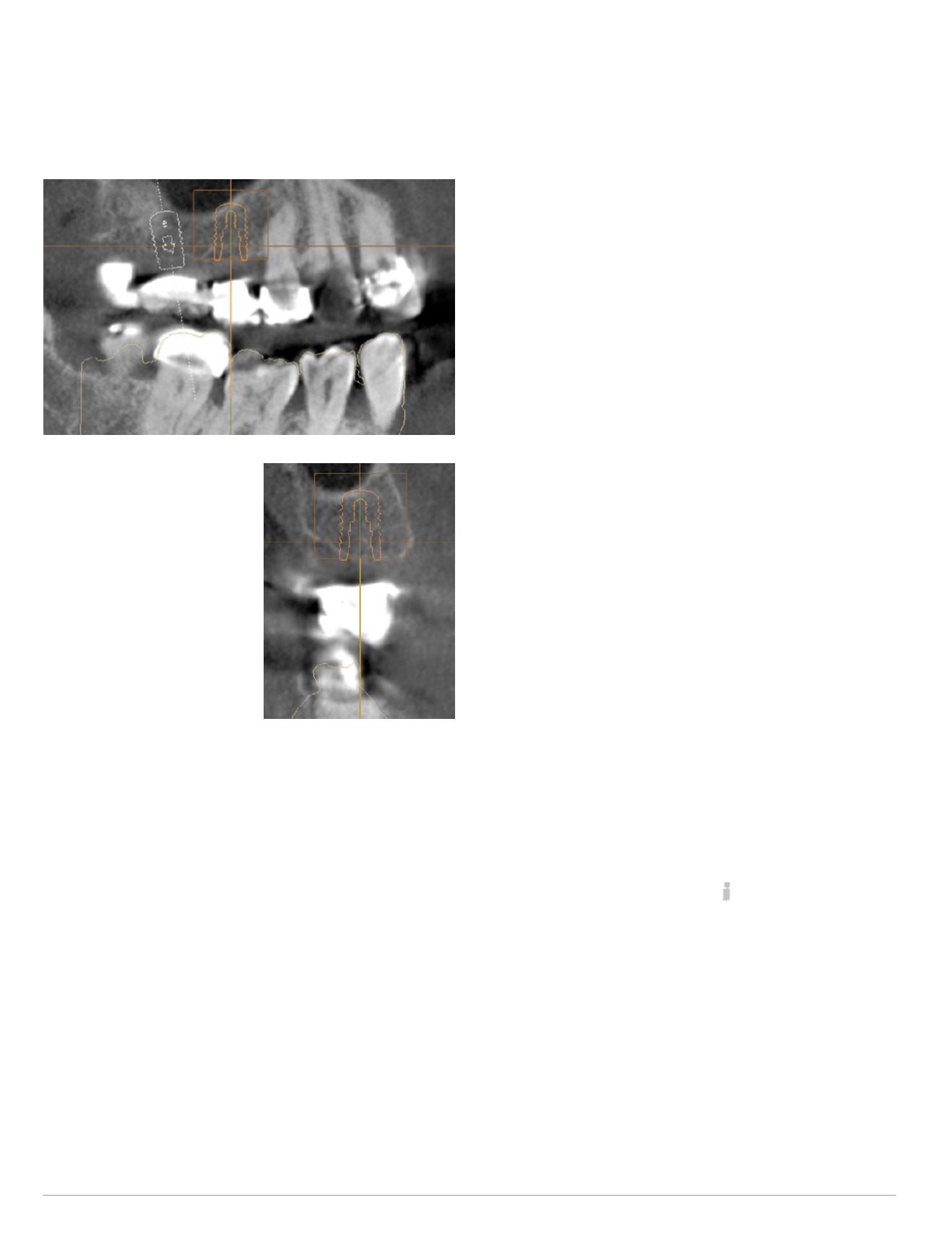
replace an ill-fitting bridge and missing teeth #3 and #4 without
involving the maxillary sinus (Figs. 26-27). Computer-assisted
surgical guides will make this a safe, positive patient experience.
Neither a 2-D panoramic X-ray, nor a 2-D periapical X-ray, would
yield this information, requiring me to recommend her to an oral
surgeon. Thus, another advantage of having this information at my
disposal is keeping all the treatment in one location.
PHASE III
1.
Replacement of ill-fitting bridge #2 through #5, with
individual crowns on teeth #2 and #5.
2.
Guided implant placement for teeth #3 and #4.
3.
Restore implants for teeth #3 and #4.
DIGITAL WORKFLOW AND GUIDED IMPLANT SURGERY
A SICAT OptiGuide is derived from a completely digital workflow
replicating the patient’s surface anatomy and surrounding anatom-
ical structures from a CBCT scan merged with a CEREC optical
impression. These templates used during guided implant surgery
| | |
R A M I R E Z
Fig. 26 (above): Tangential view
UR virtual implants #3 and
#4, periapical pathology #6
Fig. 27 (right): Cross-sectional
view virtual implant #4


