5
All onlays were adhesively bonded to the preparations using
All-Bond 2 (Bisco) and Variolink II (Ivoclar). An Instron was used
to load the onlays in the central fossa to failure and measure the
mean fracture resistance. There was a significant difference in the
fracture resistance between the materials with IPS e.max onlays
being significantly stronger than the IPS Empress onlays. This is
not a surprising finding, considering the flexural strength of IPS
e.max is approximately two to three times greater than that of IPS
Empress. However, there was no reported difference in fracture
resistance between 1 mm and 2 mm thickness onlays for either
material.
This may be considered a surprising outcome in view of the
strength differences between the materials, and considering that
reducing the thickness from 2 mm to 1 mm potentially reduced
the ceramic strength by a factor of four. This unexpected outcome
might be explained by the supportive potential of adhesively
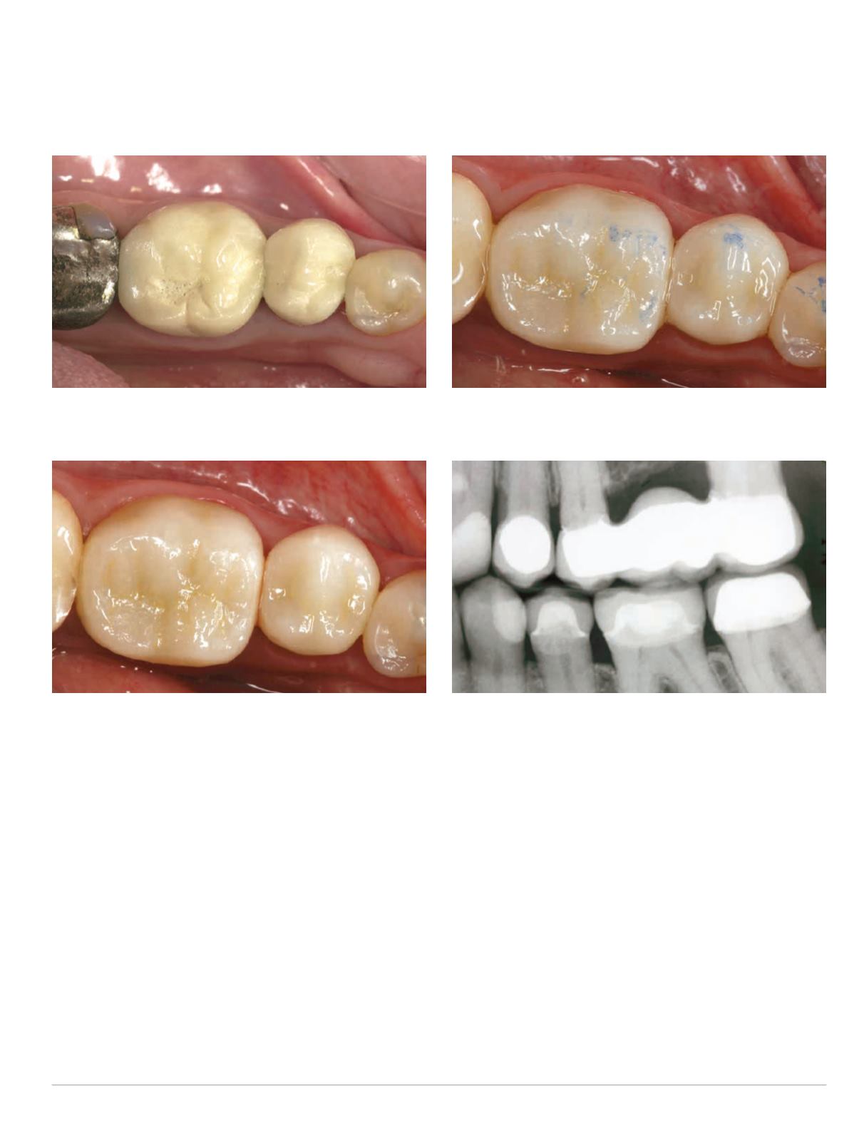
Fig. 1: Pre-operative view of PFMcrowns #19 and #20 with worn
occlusal veneer porcelain
Fig. 2: One-year recall of e.max CAD crowns #19 and #20
Fig. 3: One-year recall of e.max CAD crowns #19 and #20, with
occlusal contacts marked
Fig. 4: BWXR of left side at one-year recall examination #19 and #20
bonding the onlays to the underlying tooth structure.
Another in vitro study assessed the effect of wall thickness on
the fracture loads of monolithic lithium disilicate molar crowns
(Seydler, et al, 2014). Standardized crown preparations were
completed on 48 extracted molar teeth. The crown preparations
had different occlusal and axial wall thicknesses (0.5, 1.0 and 1.5
mm). Lithium disilicate (e.max CAD; Ivoclar) crowns were fabri-
cated with the CEREC system (Sirona) and adhesively luted to the
extracted teeth with Multilink (Ivoclar). Each group of 16 crowns
had the same occlusal and axial wall thickness. Eight crowns were
loaded immediately and the other eight crowns underwent artifi-
cial aging with thermocycling and chewing simulation. All crowns
were loaded until fracture on one cusp with a tilt of 30° to the
tooth axis in a universal testing machine. There was no signifi-
cant difference in the fracture loads for crowns that had 1.0 mm
and 1.5 mm wall thickness. Crowns with 1.0 mm and 1.5 mm wall


