55
tissue from the etchant and/or self-etching adhesive (Fig. 17).
It also helped to facilitate clean up. A universal bonding agent
(Adhese Universal) was then scrubbed onto the built-up prepa-
ration for 30 seconds, air thinned, and light-cured for 10 seconds
using an LED curing light (Bluephase Style).
An esthetic light- and dual-curing cement (Variolink Esthetic)
in shade DC Warm was placed into the restoration, after which
it was seated. This cement was selected based on its ability to
facilitate easy clean up of excess cement, shade/color stability as
a result of the patented Ivocerin, and high bond strengths. Dental
professionals can easily remove excess material after successful
pre-polymerization with light. The cement’s Viscosity Controller
contributes to the material’s good flow properties and stability, so
it can be easily extruded from the syringe.
After the restoration was seated, a Butler rubber tip was used to
remove immediate excess cement, and the restoration was tack-
cured from the buccal and lingual aspects for three seconds each.
The now gel-like excess cement was removed (Fig. 18), and the
restoration was fully cured from all angles for 10 seconds each
(Fig. 19). The occlusion was checked and adjusted as needed,
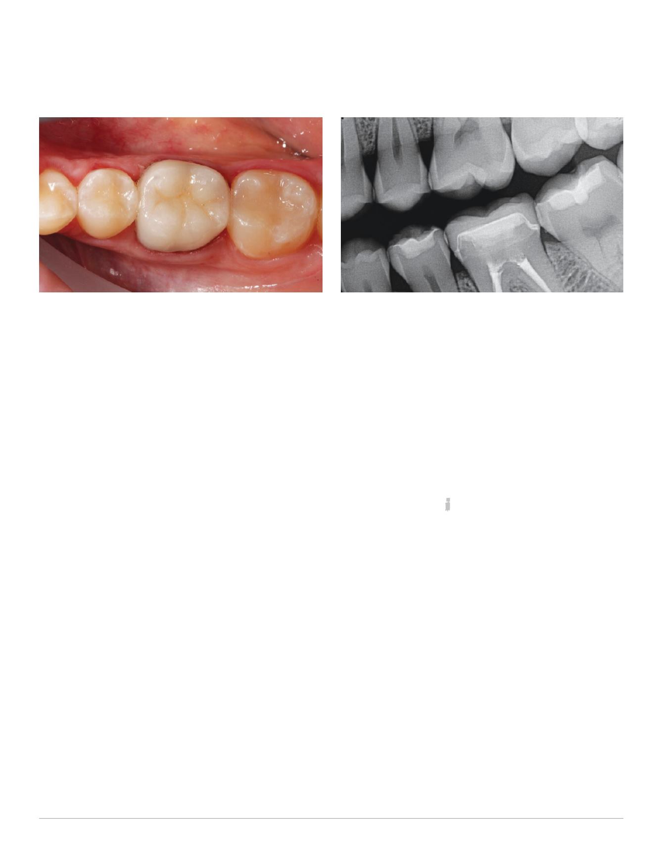
and the restoration was polished. Postoperative photographs and
radiograph were then taken to confirm complete marginal integ-
rity and removal of excess cement (Figs. 20-21).
CONCLUSION
When a combination of advanced materials and technologies are
used, patients can receive predictable indirect restorations that
demonstrate an exceptional fit and esthetics in one office visit.
In the case presented here, in-office design, milling and seating
of a lithium disilicate (IPS e.max CAD), full-coverage crown was
simplified by taking advantage of digital intraoral impression
scans, restoration design software, and luting materials that work
together to provide immediate and long-lasting bond strengths,
esthetics and durability. In particular, Variolink Esthetic light-
and dual-curing adhesive luting composite contributed to overall
esthetic cementation predictability and simplicity. Additionally,
it also promoted precise shade matching of the restoration to
the surrounding natural tooth structure and adjacent bulk-fill
composite restorations.
For questions and more information, Dr. Vasquez can be reached at
.
REFERENCES
1 MiyazakiT,HottaY,KuniiJ,KuriyamaS,TamakiY.Areviewofdentalcad/cam:current
statusandfutureperspectivesfrom20yearsofexperience.DentMaterJ.2009;28(1):44-56.
2 Christensen GJ. In-office cad/cammilling of restorations. J AmDent Assoc.
2008;139(1):83-85.
3 RekowED, Erdman AG, Riley DR, Klamecki B. CAD/CAM for dental restorations –
some of the interesting challenges. IEEE Trans Biomed Eng. 1991;38(4):314-8.
4 Liu PR. A panorama of dental cad/cam restorative systems. Compend Contin Educ Dent.
2005;26(7):507-8, 510, 512 passim, quiz 517, 527.
5 FasbinderDJ.Thecerecsystem:25yearsofchairsidecad/camdentistry.JAmDentAssoc.
2010;141 Suppl 2:3S-4S.
6 Stutes RD. The history and clinical application of a chairside cad/camdental restoration
system. Shanghai Kou Qiang Yi Xue. 2006;15(5):449-55.
7 Culp L, McLaren EA. Lithiumdisilicate: the restorative material of multiple options.
Compend Contin Educ Dent. 2010;31(9):716-20, 722, 724-5.
8 Manso AP, Silva NR, Bonfante EA, et al. Cements and adhesives for all-ceramic
restorations. Dent Clin North Am2011;55(2):311-32, ix.
9 McComb D. Adhesive luting cements—classes, criteria, and usage. Compend Contin
Educ Dent. 1996;17(8):759-62; 764 passim; quiz 774.
10 Rickman LJ, Satterthwaite JD. Considerations for the selection of a luting cement.
Dent Update. 2010;37(4):247-8, 251-2, 255-6 passim.
Fig. 20: Postoperative occlusal view of the bulk-filled restorations and
the lithium disilicate CAD/CAMcrown
Fig. 21: Postoperative radiograph demonstrating marginal integrity
and seal among all bulk-filled restorations and the lithium
disilicate CAD/CAMcrown


