41
DISCUSSION
The transcrestal sinus floor elevation technique has been shown to
be a predictable approach, enabling implant placement in maxil-
lary posterior sites with a mild-to-moderate vertical alveolar ridge
deficiency due to the pneumatization of the Maxillary sinus. The
above case example outlines a predictable technique to combine
this approach with computer-guided implant surgery to place
the implant in a three-dimensionally correct prosthetic position
thereby ensuring a long-term successful outcome.
An argument can be made that a socket bone graft for ridge pres-
ervation shouldhave beenperformed in this case at the time of tooth
extraction. This is certainly a viable treatment option. However,
since there was a relatively adequate, albeit limited, bone height to
the maxillary sinus in this case post-extraction and, since the bone
was very dense in this particular patient, the decision was made not
to perform any bone grafting at the time of extraction.
This would allow unimpeded soft and hard tissue healing to
take place, taking into account that any mild to moderate residual
alveolar ridge defects could be grafted very predictably at the
second stage implant placement surgery.
In addition, another argument can bemade that computer-guided
implant surgery is not very beneficial in these type of cases since
it only allows for minimal guidance in the residual 4 mm to 5 mm
of bone below the maxillary sinus. However, one should realize
that guided implant surgery allows for the maximum utilization of
the existing osseous support in accordance with the prosthetically
driven implant treatment plan. In addition, since the bone quality in
the posterior Maxilla is oftentimes
very soft (Type IV bone), free-hand
implant osteotomy preparation and
implant placement in these type of
cases with only 4 mm to 5 mm of
residual host bone can very easily
lead to inaccuracies in implant posi-
tioning and placement.
Computer-guided implant surgery
helps to avoid these inaccuracies and
ensures accurate implant positioning
and placement through the restric-
tion of the surgical guide.
Dr. Boltchi would like to thank Dr. Mike Rogers (Arlington, Texas) for
the restorative treatment provided for the patient in this case example.
For questions and more information, Dr. Boltchi can be reached at
.
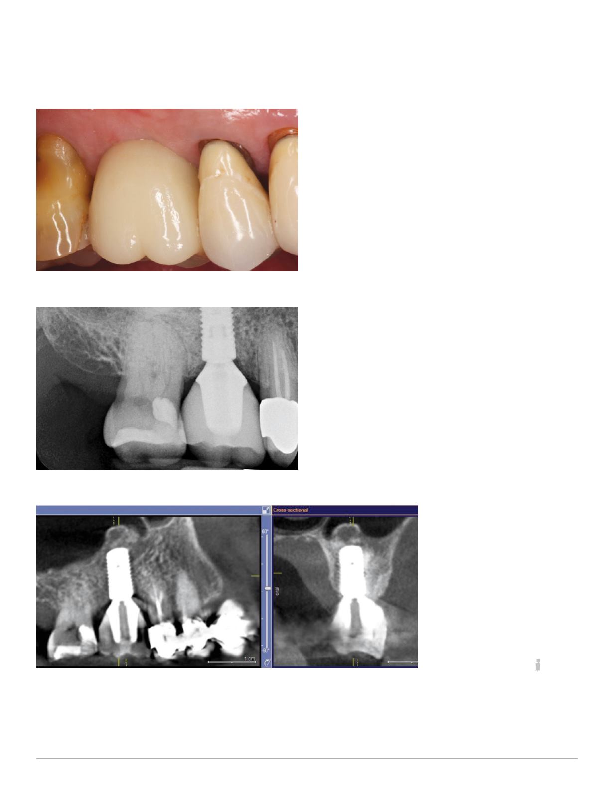
Fig. 21: Facial view of final implant restoration #3
Fig. 22: Periapical radiograph of final implant restoration
Fig. 23: Postoperative CBCT scan


