29
The PRF plugs were cut into
smaller pieces and several of
these smaller pieces were mixed
with a cortico-cancellous bone
allograft (MinerOss, BioHo-
rizons). The resulting space/
pouchbetween the residual alve-
olar ridge and the SonicWeld Rx
membrane was then filled with
this bone graft mixture, thereby
forming a three-dimensionally
stable and rigid reconstructive
unit (Fig. 10). A pericardium
membrane (Copi-Os, Zimmer)
was placed over the SonicWeld
Rx membrane and bone graft
to cover the bone graft crest-
ally (Fig. 11), and several PRF
membranes were then layered
over this pericardiummembrane
(Fig. 12). A periosteal releasing
incision was performed buccally
in order to advance the buccal
flap coronally and to obtain a
tension-free primary wound
closure (Fig. 13).
The patient was re-evaluated
clinically and radiographically
after an uneventful healing
period of six months (Figs.
14-15). Based on this initial
clinical and radiographic evalu-
ation, it was concluded that the
bone graft was well consoli-
dated. The patient was then
scheduled for the second-stage
implant placement surgery. A
cone beam CT radiographic
evaluation was performed with
the Sirona Orthophos XG3D
CBCT machine, a laboratory
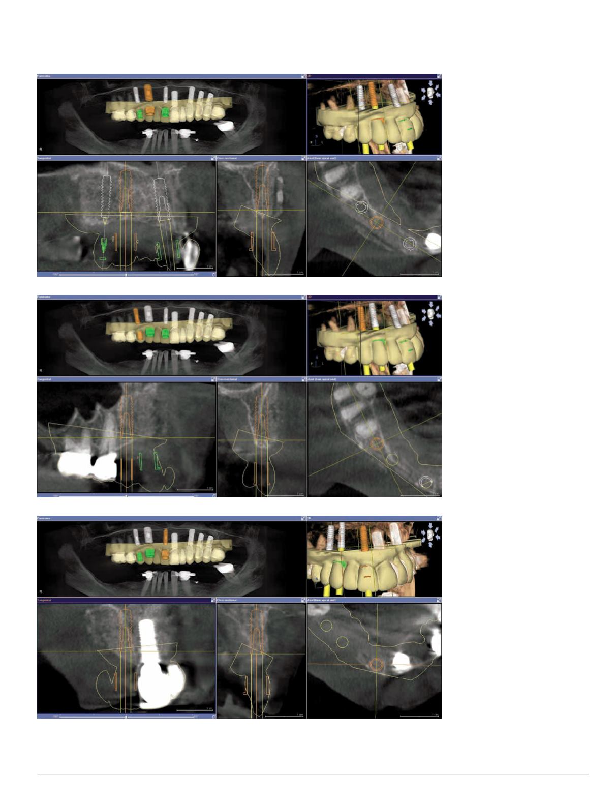
Figs. 16-18: (Top to bottom)
CBCT implant treatment plan
sites #5, #6 and #8


