31
OptiGuide apply to the surgical Guide. In the case example above,
however, we were able to obtain an accurate merging of the CAD/
CAM data with the CBCT data despite the abundance of pre-
existing metallic restorations, and the resulting Digital Guide was
therefore very accurate. This may, however, not always be the case
in situations with pre-existing metallic restorations due to the
scatter artifact caused by these restorations, and should therefore
be evaluated on a case-by-case basis.
SICAT has tested the accuracy of the Digital Guide with the
Stratasys Objet Eden 260V printer, and it is recommended to use
a professional-grade 3-D printer with a similar accuracy for the
fabrication of these guides. In the case example above, we sent
the Digital Guide .STL CAD file to a professional printing center
(Dominion Milling Center, Virginia), where the guide was printed
with a 3-D Systems ProJet 3000 printer. In addition, it should be
noted that these guides must be printed with printers utilizing
materials that are FDA-approved for intraoral use.
In our limited experience thus far, the SICAT Digital Guide
has proven to be very accurate. However, one has to pay atten-
tion to the accuracy of the 3-D printers utilized for the fabrication
of these guides and to the careful insertion of the corresponding
guided surgery sleeves as needed. The Digital Guide should there-
fore not be regarded as a replacement for the Classic Guide or the
OptiGuide and, to ensure the highest level of accuracy, should only
be utilized according to its intended indication.
For questions and more information, Dr. Boltchi can be reached at
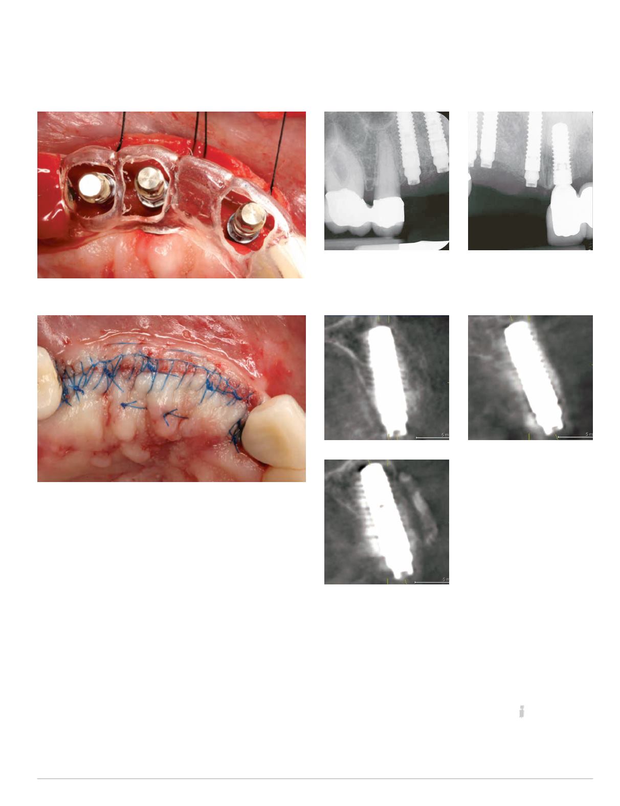
Fig. 22: Restoratively correct implant positioning, sites #5, #6 and #8
Fig. 23: Primary wound closure for submerged implant healing
Fig. 24: Immediate post-implant
placement, periapical
radiograph sites #5-6
Fig. 25: Immediate post-
implant placement periapical
radiograph sites #6-8
Figs. 26-28: Postoperative cross-
sectional CBCT view, sites #5
(above left), #6 (left) and #8
(above)


