27
CASE STUDY
This patient is a 73-year-old female, who was referred for dental
implant evaluation in sites #5-8 after failure of a long-span, fixed
partial denture#3-8due to fracture of the anterior abutment tooth#8.
The initial clinical and periapical radiographic evaluation revealed
a restoratively hopeless tooth#8, whichwas fracturedhorizontally at
the gingival level, and an edentulous alveolar ridge in sites #5-7 post
sectioning and removal of the failed fixed partial denture (Figs. 1-4).
Sincetheinitialclinicalevaluationrevealedonlyamild-to-moderate
alveolar ridge deficiency in sites #5-8, a preliminary treatment plan
was devised consisting of extraction of tooth #8 and immediate
implant placement in sites #5, #6 and #8 with simultaneous bone
grafting. This treatment plan was to be confirmed or modified based
on a cone beam computerized tomographic (CBCT) evaluation. The
cone beam CT radiographic evaluation, performed with the Sirona
Orthophos XG3D CBCT, revealed a severe horizontal alveolar ridge
defect in sites #5-7, and a thin buccal alveolar plate in site #8, neces-
sitating a staged alveolar ridge reconstruction prior to implant place-
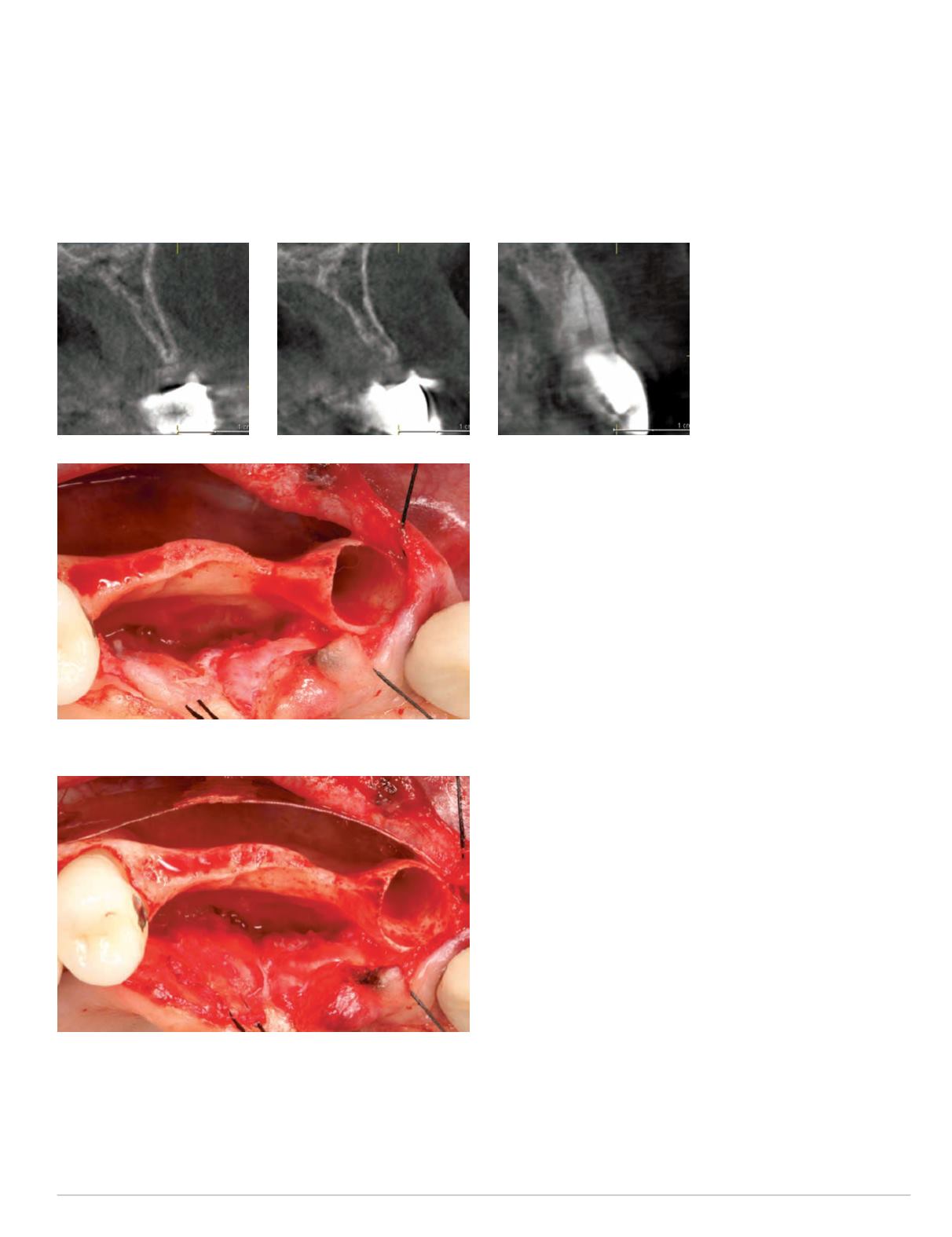
ment and amodification of the original treatment plan (Figs. 5-7).
The first-stage surgical procedure was performed under intra-
venous conscious sedation and local anesthesia, and venous blood
was collected to obtain a Platelet Rich Fibrin (PRF) concentrate
via a centrifuge system. The hopeless tooth #8 was extracted and
buccal and palatal full-thickness flaps were reflected, revealing
an extended and severe horizontal alveolar ridge defect in sites
#5-7, as well as a very thin buccal alveolar plate in site #8. This
confirmed the CBCT findings (Fig. 8).
A 50 mm x 20 mm SonicWeld Rx membrane was trimmed,
adapted to the alveolar ridge defect and welded with the Sonic-
Welder Rx sonotrode to the heads of SonicWeld Rx Pins. These
had been previously placed buccally into the alveolar bone at the
outer boundaries of the alveolar ridge defect (Fig. 9).
Fig. 5-7 (left to right):
Preoperative cross-sectional
CBCT view, sites #5, #6
and #8
Fig. 8: Sites #5-8 after flap reflection and extraction of tooth #8
Fig. 9: Adaptation of SonicWeld membrane sites #5-8


