30
|
CERECDOCTORS.COM
|
QUARTER 3
|
2015
| | |
B O LT C H I
wax-up for sites #5-8 was scanned with the CEREC Omnicam,
and the corresponding CAD/CAM data was exported as an .SSI
file and imported into the Galaxis software. Once there, it was
merged with the CBCT data. This CEREC-Galileos integration
workflow was then utilized to plan a restoratively-driven implant
placement in the Galaxis implant treatment planning software in
sites #5, #6 and #8 (Figs. 16-18).
The treatment planning data was sent to SICAT (in Bonn,
Germany) for the design of a Digital Guide. Upon designing the
surgical guide, SICAT uploaded the CAD file of the surgical guide
design as a .STL file to the SICAT portal. This .STL file was then
downloaded from the SICAT portal and sent to a professional 3-D
printing center (Dominion Milling Center, Virginia) for the fabri-
cation of the actual surgical guide. Upon receipt of the printed
surgical guide, original Straumann guided-surgery sleeves were
placed into the surgical guide, thereby completing the fabrication
of the Digital Guide (Fig. 19).
The second-stage surgical procedure was performed under
intravenous conscious sedation as well as local anesthesia. A
minimal bucco-lingual crestal flap reflection was performed
to expose the reconstructed alveolar ridge, and three implants
(Straumann 3.3 x 12mmRoxolid bone-level implants) were placed
via a guided approach in sites #5, #6 and #8 (Figs. 20-21).
The implants in sites #6 and #8 were placed via a fully guided
approach through the SICAT Digital Guide, whereas the implant
in site #5 was placed freehand after guided osteotomy preparation
with the Digital Guide. All implants were placed in restoratively
correct positions according to the preoperative treatment plan
(Fig. 22). Primary flap closure was then obtained with contin-
uous and interrupted 5.0 Polypropylene sutures to allow for a
submerged and undisturbed implant healing (Fig. 23).
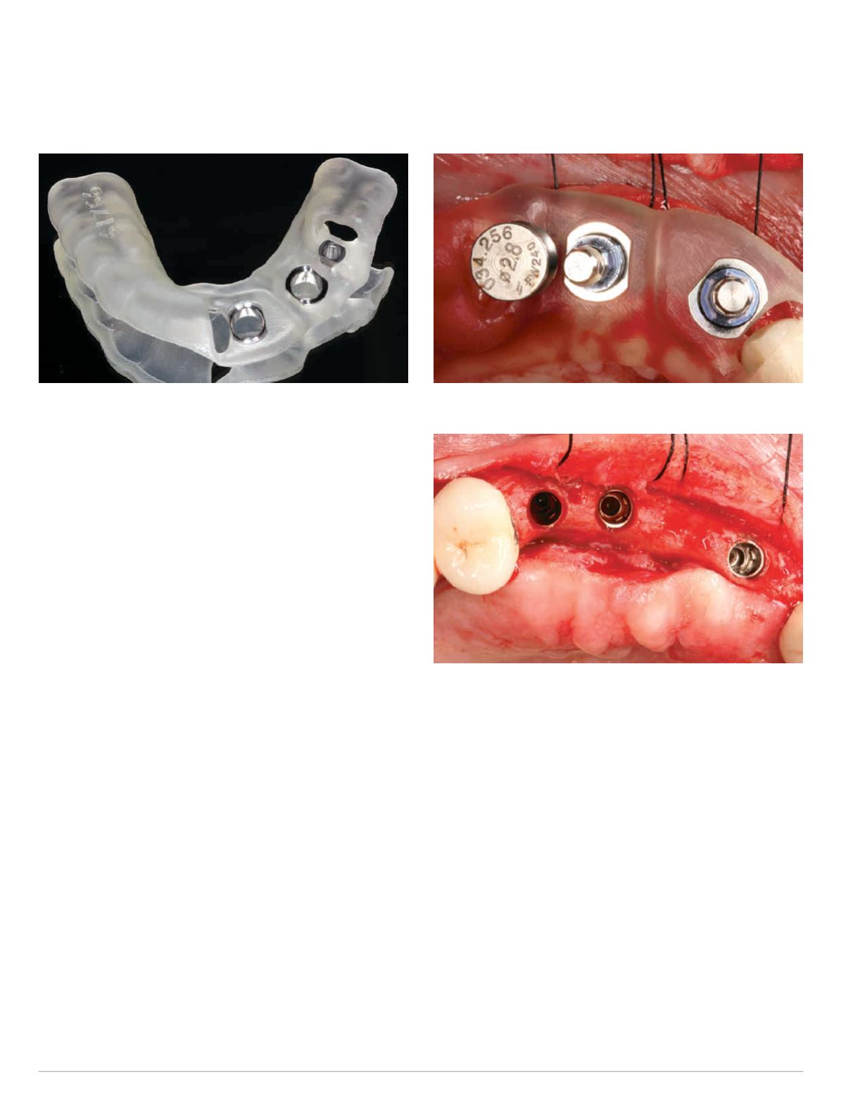
Fig. 19: SICAT Digital Guide
Fig. 20: Guided implant placement with SICAT Digital Guide
Fig. 21: Occlusal view after implant placement sites #5, #6 and #8
The immediate postoperative periapical and CBCT radiographs
confirmed the accuracy of the implant positioning according to
the preoperative plan (Figs. 24-28).
DISCUSSION
The SICAT Digital Guide is a welcome new addition to SICAT’s
surgical guide options for guided implant surgery. The main
advantage of the Digital Guide is a faster turnaround time and a
reduced cost as compared to the Classic Guide or OptiGuide.
SICAT will typically complete the surgical guide design and
upload the corresponding .STL file to the SICAT portal within two
business days. This is significantly fewer than the 10 business days
required for a Classic Guide and the six business days required for
an OptiGuide.
The workflow for the Digital Guide is identical to the workflow
for the OptiGuide, and typically the same restrictions of a minimal
amount of pre-existing metallic restorations that apply to the


