44
|
CERECDOCTORS.COM
|
QUARTER 3
|
2015
| | |
S K R A M S TA D
How do we know if the tissue will
predictably fill in the gap? Obviously,
biology controls this process, but from
a design perspective, you have to
remember that the tissue is “retracted”
in the prep image. You need to design
the apical-most aspect of the contact
point to the un-retracted tissue in
the Biogeneric Copy folder.
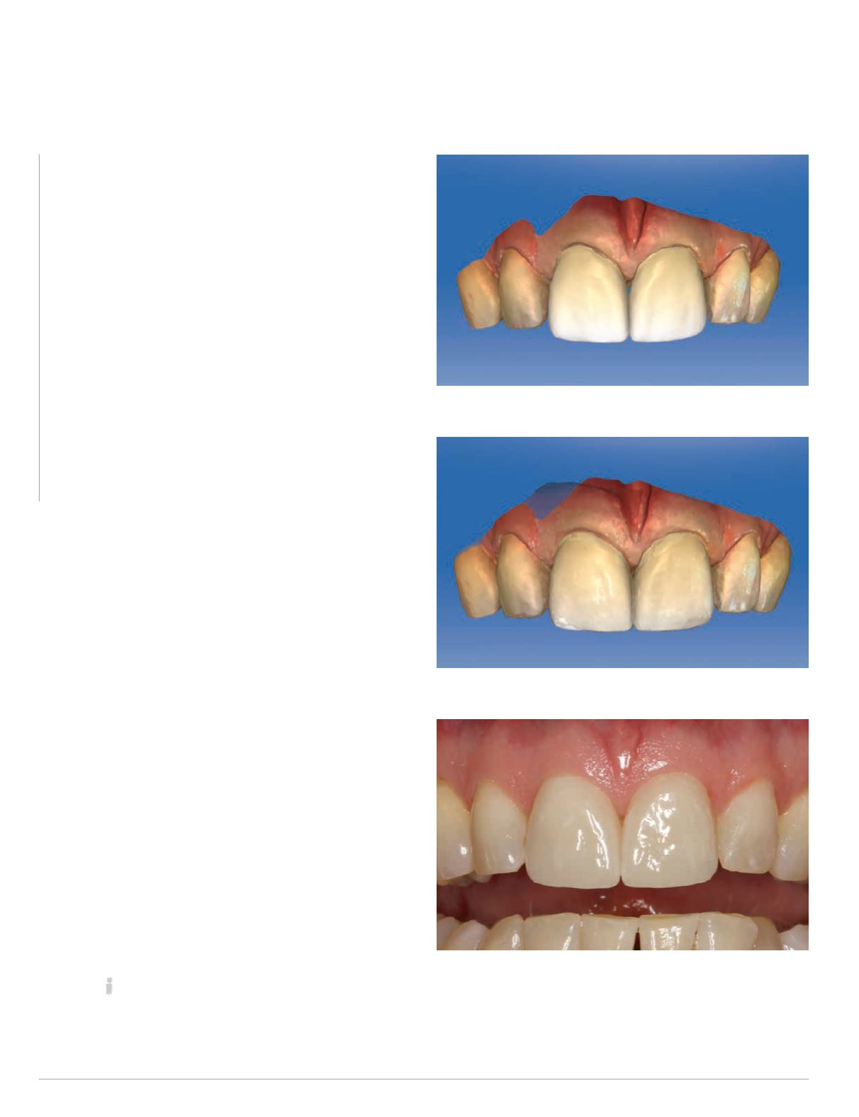
Fig. 18: Final design, #8 and #9
Fig. 19: Checking tissue with Biocopy
Fig. 20: Final restorations, seated
see with the final design of the restorations (Fig. 18), the teeth have
remained symmetrical throughout the entire design process and I
was able to create a proper midline that was long and flat.
There is one last critical design trick that must be understood
for proper form of central incisors. That is the length of the
contact. You can see from my final design picture that there is a
black triangle present. I could quite easily close that contact from
the lingual if it was needed, but I did not.
How do we know if the tissue will predictably fill in the gap?
Obviously, biology controls this process, but from a design
perspective, you have to remember that the tissue is “retracted”
in the prep image. You need to design the apical-most aspect
of the contact point to the un-retracted tissue in the Biogeneric
Copy folder.
If we overlay the Biocopy folder transparent over the design
(Fig. 19), you can see that the tissue fills in the slight gap left in my
final design, and I would expect it to fill in intraorally as well. If
you look at the final photos (Fig. 20), it did just that.
Hopefully this article helped you understand symmetry with
central incisors and, more importantly, how to maintain the
symmetry throughout the design process to get proper form and
beautiful final results. There is much more to talk about regarding
anterior form, function and esthetics, and I look forward the next
discussion.
For questions and more information, Dr. Skramstad can be reached at
.


