20
|
CERECDOCTORS.COM
|
QUARTER 2
|
2016
C A S E S T U D Y
| | |
B Y FA R H A D E . B O LT C H I , D . M . D . , M . S .
the cerec digital implant dentistry workflow provides a completely integrated experience
incorporating
all aspects of surgical and restorative implant dentistry. The incorporation of in-house milling of surgical guides and in-house
fabrication of custom abutments and screw-retained crowns has been instrumental in this regard.
Immediate Implant Placement
With CEREC Guide 2
A Step-by-step Overview of This Fully Integrated Digital Workflow
The recent introduction of CEREC Guide 2 — coupled with
innovative restorative components for the CEREC digital implant
dentistry workflow such as Straumann’s Variobase for CEREC,
Ivoclar’s Telio CAD provisional Implant Solutions block, and
Ivoclar’s e.max Hybrid Abutment block — significantly improves
this digital implant dentistry workflow. It enables clinicians to
performvery precise and efficient in-house digital implant therapy
with the CEREC CAD/CAM system. This article will provide a
step-by-step overview of this digital workflow.
scanning with the CEREC Omnicam. A virtual restoration was then
designed in theCERECChairside software4.4, and the corresponding
CAD/CAM data was exported into the Galileos Implant treatment-
planning softwarewhere it wasmergedwith the CBCT scan.
The Galileos Implant treatment planning software was utilized
to plan a Straumann Bone Level Tapered SLActive Roxolid RC 4.1
mm x 14 mm Implant in site #9 (Fig. 4). The treatment-planning
data was then exported from the Galileos Implant treatment-
planning software, and imported back into the CEREC Chairside
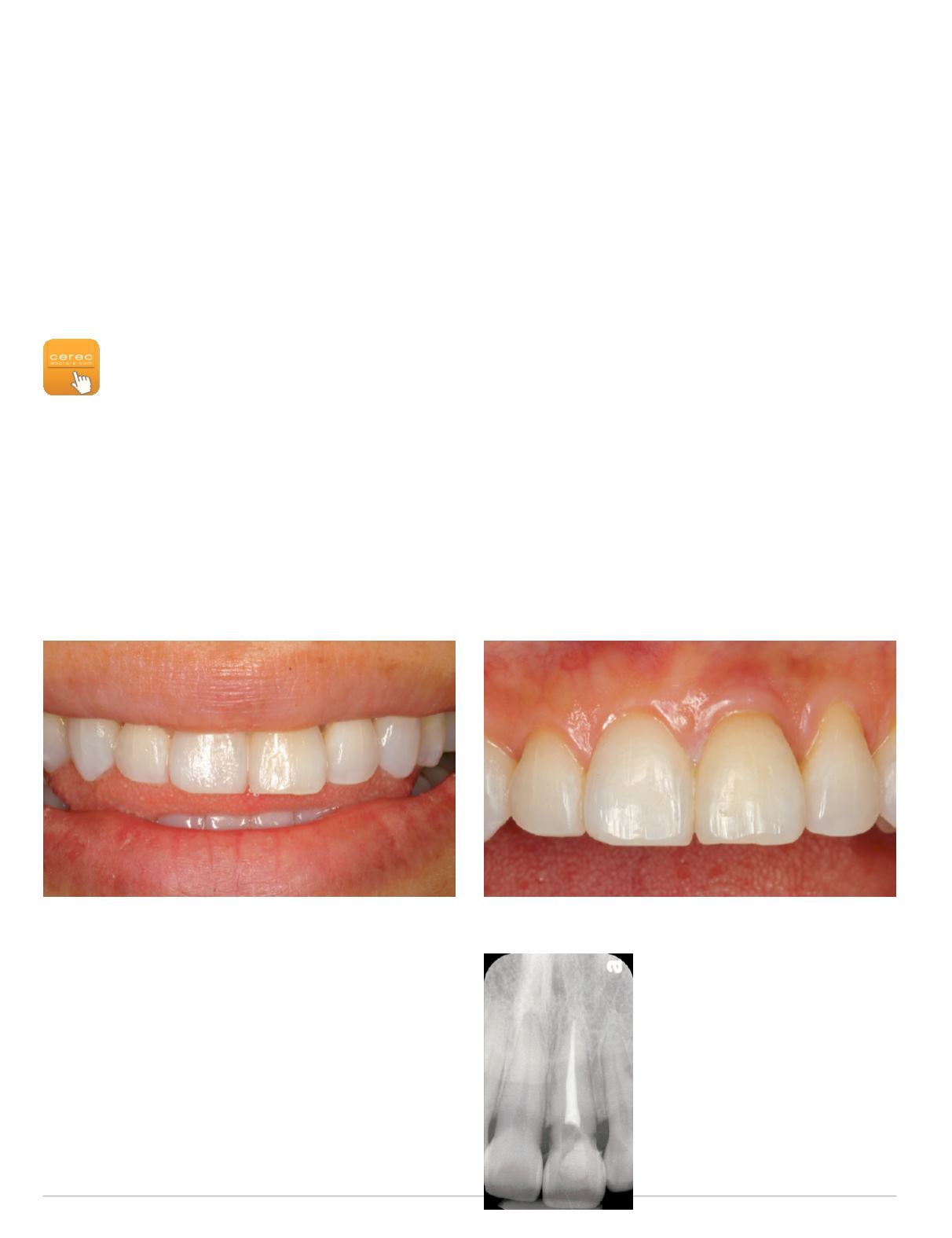
Fig. 1: Preoperative smile view
CASE STUDY
The 32-year-old female patient presented for implant therapy to
replace a fractured and hopeless tooth #9. She had a non-contrib-
utory medical history. The initial clinical and periapical radio-
graphic evaluation revealed a medium-high lip line, a relatively
thin periodontal biotype and an oblique crown/root fracture of
tooth #9 (Figs. 1-3).
A cone beam CT radiographic evaluation was performed with the
Sirona Orthophos XG3D CBCT machine, and a digital impression
of the patient’s maxillary and mandibular arches was obtained via
Fig. 2 (above): Preoperative retracted facial view
Fig. 3 (left): Preoperative
periapical radiograph
of fractured tooth
site #9


