30
|
CERECDOCTORS.COM
|
QUARTER 2
|
2016
KEY TAKEAWAY
I propose that you always take the Biocopy scan as a back-up
folder even though that may not be your design method. Superim-
posing your Biocopy folder over your design will not only help you
with the position of the papillae but also with other details like the
midline, incisal edge position and line angles.
For questions and more information, Dr. Patel can be reached at
.
came in after a skiing accident and had fractured her upper centrals
(Fig #3). Both teeth had fractures running into the cingulum with
exposed pulp. Both teeth were treated with endo and nowwe had to
do crowns on both. Again, in this case, as the shape of the teeth was
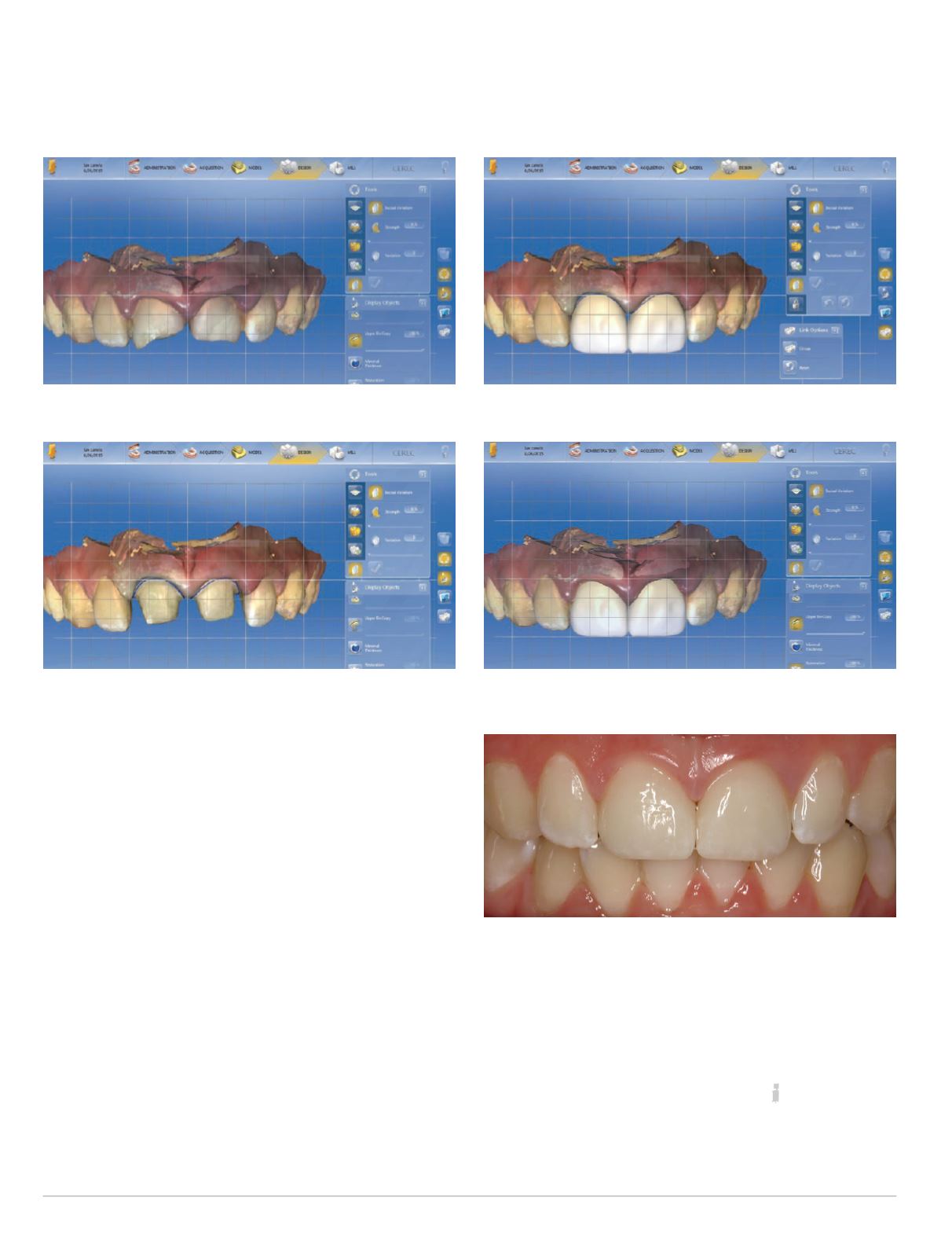
not desirable, I chose Biocopy Individual to designmy case. But I also
scanned the pre-operative condition as my Biocopy folder (Fig #4).
Both teethwere prepped for crowns and scanned in the prepped
folder (Fig #5). This is my final design after using a few tools and
the grid to make sure my centrals are symmetrical (Fig #6). As you
can see, there is open space between #8 and #9 which most of us
would be inclined to close by adding some porcelain.
Here iswhereIwouldsuggest that youoverlayyourBiocopy folder
over the design. When you do this, you can see the original position
of the papillae in comparison to your designed restoration (Fig #7).
Remember, in the prep folder, the gingiva has been pulled back due
to a cord being packed and are not where they usually belong. Once
you make sure that the original position of the papillae will fill that
open space, you no longerwill close that gap andwill finishupwith a
result where the papillae are not blunted and the contacts don’t look
too long — like they did on the earlier case (Fig #8).
Fig. 4: Biocopy model
Fig. 5: Preparations
Fig. 6: Final design
Fig. 7: Biocopy overlayed on proposals for tissue position
Fig. 8: Final result
| | |
PAT E L


