36
|
CERECDOCTORS.COM
|
QUARTER 1
|
2016
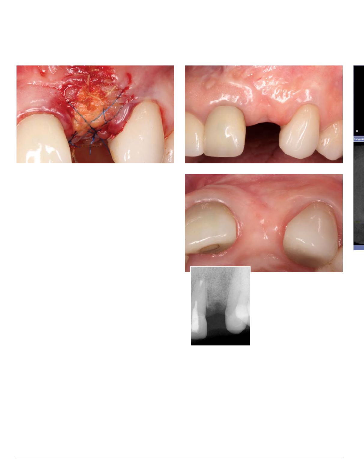
tissue graft in place, intentionally leaving the connective tissue
graft exposed (Fig. 11). The non-resorbable sutures were removed
after two weeks and the Cytoplast membrane, which had become
exposed after three weeks, was removed four weeks after its initial
exposure or seven weeks after the initial reconstructive surgery.
The second treatment phase was performed after an uneventful
healing period of 6 months to allow for complete soft and hard tissue
healing of the soft and hard tissue grafts in site #10. The clinical and
periapical radiographic re-evaluation revealed a completely healed
site with an excellent result of the previously performed hard and soft
tissue reconstruction (Figs. 12-14). Basedon this initial evaluation itwas
concluded that the bone graft was well consolidated and the patient
was scheduled for the second stage implant placement surgery.
A cone beamCT radiographic evaluationwas performedwith the
Sirona Orthophos XG3D CBCT machine, a digital intra-oral scan
was obtained with the CEREC Omnicam, a virtual restoration was
designed in the CEREC chairside software, and the corresponding
CAD/CAM data was exported as a .ssi file and imported into the
Galaxis software, where it was merged with the CBCT data. This
CEREC-Galileos integration workflow was then utilized to plan
a restoratively-driven implant placement in the Galileos Implant
treatment planning software in site #10 (Fig. 15).
The treatment planning datawas sent to SICAT in Bonn, Germany
for the design of a Digital Guide. Upon designing the surgical guide,
SICAT uploaded the CAD file of the surgical guide design as a .stl
file to the SICAT portal. This .stl file was then downloaded from
the SICAT portal and sent to a professional 3-D printing center
(Dominion Milling Center, Virginia) for the fabrication of the actual
surgical guide. Upon receipt of the printed surgical guide, an original
Straumann guided surgerymaster sleevewas placed into the surgical
guide, thereby completing the fabrication of the Digital Guide.
The second stage surgical proce-
dure was performed under intrave-
nous conscious sedation and local
anesthesia as well. Implant place-
ment in site #10 was accomplished via a flapless guided approach.
The SICAT Digital Guide and the Straumann Guided Surgery System
were utilized to prepare the guided implant osteotomy according
to the virtual treatment plan in the Galileos Implant software and a
Straumann 3.3 mm X 14 mm SLActive Roxolid Bone Level implant
was placed through the Digital Guide in a fully guided fashion in
the restoratively correct and pre-planned position (Figs. 16-17). The
implant achieved excellent primary stability and was placed in a flap-
less approachwithin the confines of the osseous alveolar housing.
Since the implant achieved excellent primary stability and in order
to start developing the implant tissue transition zone a decision was
| | |
B O LT C H I
Fig. 11: Sutured surgical site after insertion of connective tissue graft
Figs. 12-14: Facial (top) and occlusal
(above) clinical views and periapical
radiograph (left) six months post
bone graft


