38
|
CERECDOCTORS.COM
|
QUARTER 1
|
2016
| | |
B O LT C H I
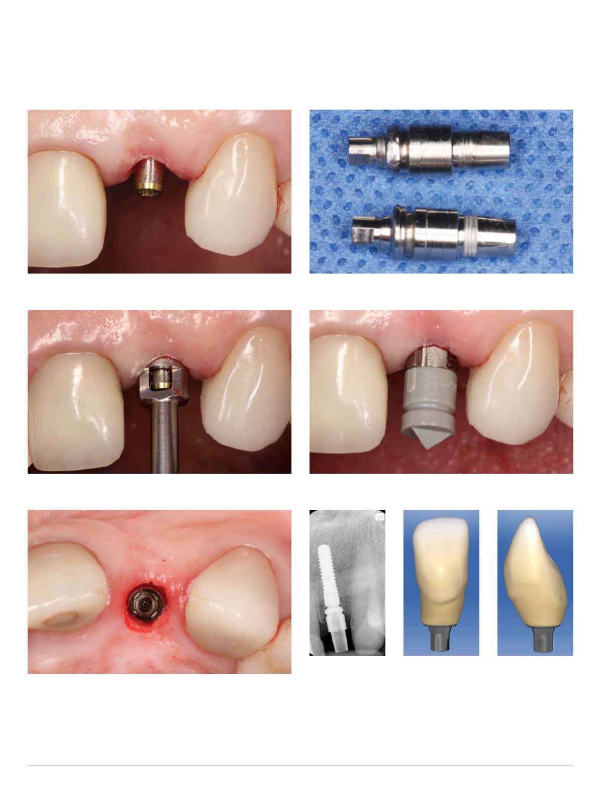
Fig. 18: Insertion of guided bone profiling pin
Fig. 19: Guided soft tissue and bone profiling
Fig. 20: Clinical view after completed guided soft tissue and bone
profiling
Fig. 21: ScanPost shoulder diameter reduction
Fig. 22: Clinical view of inserted ScanPost and ScanBody
Fig. 23: Periapical
radiograph of
seated ScanPost
Fig. 24: Facial
view of CEREC
restorative design
Fig. 25: Inter-
proximal view
of CEREC
restorative
design


