35
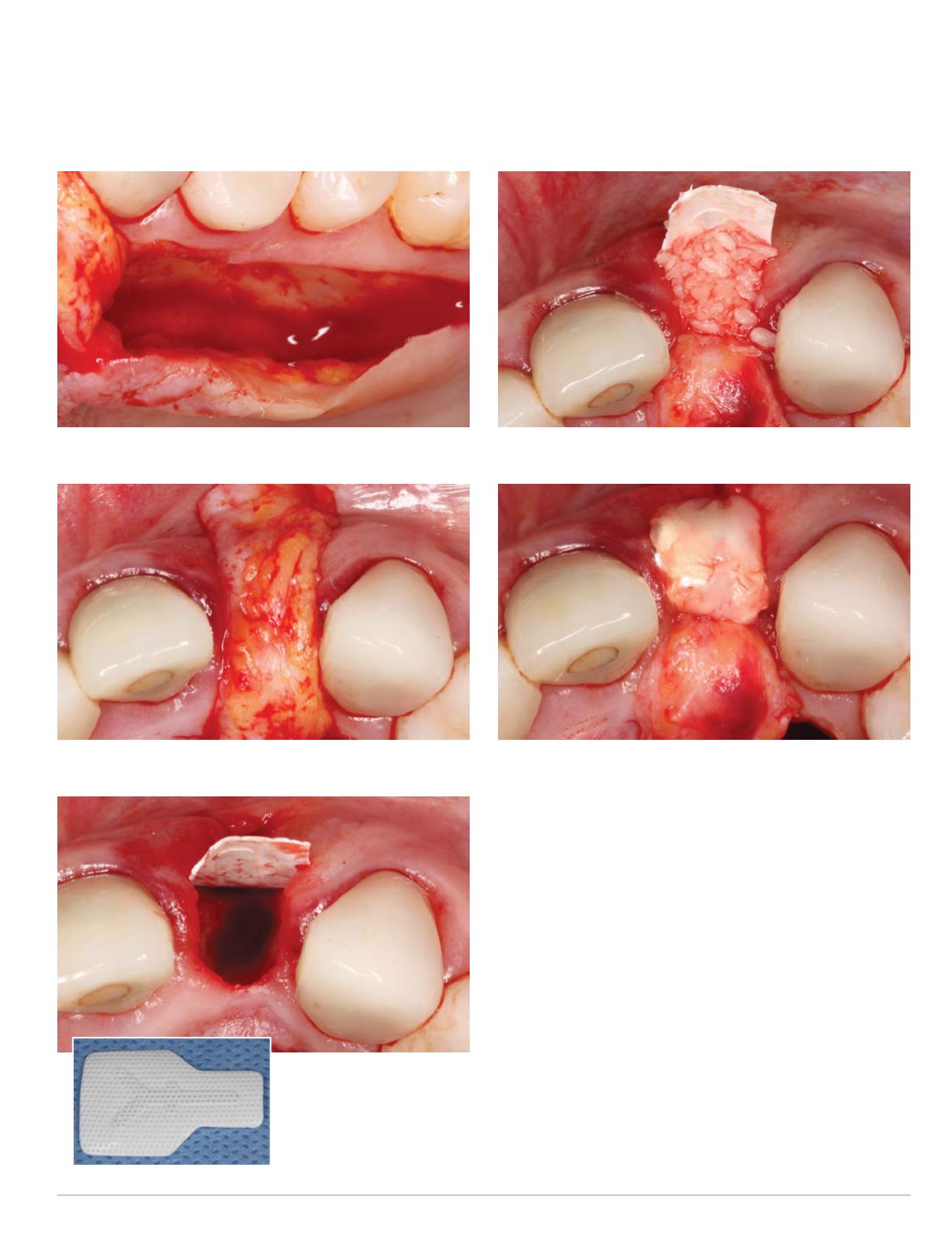
the extraction site to ensure that it could be inserted into the buccal
pouch ina completelypassivemannerwithout any tensionon the graft
(Fig.6).AtitaniumreinforceddensePTFECytoplastbarriermembrane
was then trimmed and inserted into the buccal pouch (Figs. 7-8).
Several PRF membranes were cut into small pieces and mixed
with Straumann cortico-cancellous mineralized bone allograft,
which had been reconstituted previously with the PRF growth
factor rich supernatant liquid. This bone graft mixture was inserted
into the socket in site #10 (Fig. 9), the Cytoplast membrane was
folded over the bone graft and tucked into the palatal pouch, and
a PRF membrane was inserted into the buccal and palatal pouches
covering the Cytoplast membrane (Fig. 10).
The pedunculated connective tissue graft was then rotated and
inserted into the buccal pouch over the PRF membrane covering
both the Cytoplast and PRF membranes. Various resorbable and
non-resorbable interrupted and mattress sutures were used to
approximate the wound margins and to secure the connective
Fig. 5: Palatal harvesting of pedunculated connective tissue graft
Fig. 6: Pedunculated connective tissue graft passively draped over site #10
Figs. 7-8: Cytoplast titanium
reinforced barriermembrane
(right) inserted into buccal
pouch (above)
Fig. 9: Socket grafting with Straumann mineralized allograft
Fig. 10: Cytoplast and PRFmembranes folded over bone graft


