34
|
CERECDOCTORS.COM
|
QUARTER 1
|
2016
C A S E S T U D Y
| | |
B Y FA R H A D E . B O LT C H I , D . M . D . , M . S .
sirona’s integrated digital implant dentistry workflow not only simplifies dental implant therapy and
makes it more efficient but it also adds a level of precision that surpasses other techniques.
An increasing number of dental implant manufacturers are recognizing the unique advantages of this digital workflow and
are therefore collaborating with Sirona to provide their surgical and restorative components for the CEREC CAD/CAM system. This
article will provide a case example to demonstrate Straumann’s newly introduced Variobase for CEREC.
Fully Integrated Implantology
With Straumann’s Variobase for CEREC
Sirona’s Integrated Digital Implant Dentistry Workflow
Is a Winning Combination Even in Complex Cases
CASE STUDY
This patient is a 57-year-old female
patient, who was self-referred for
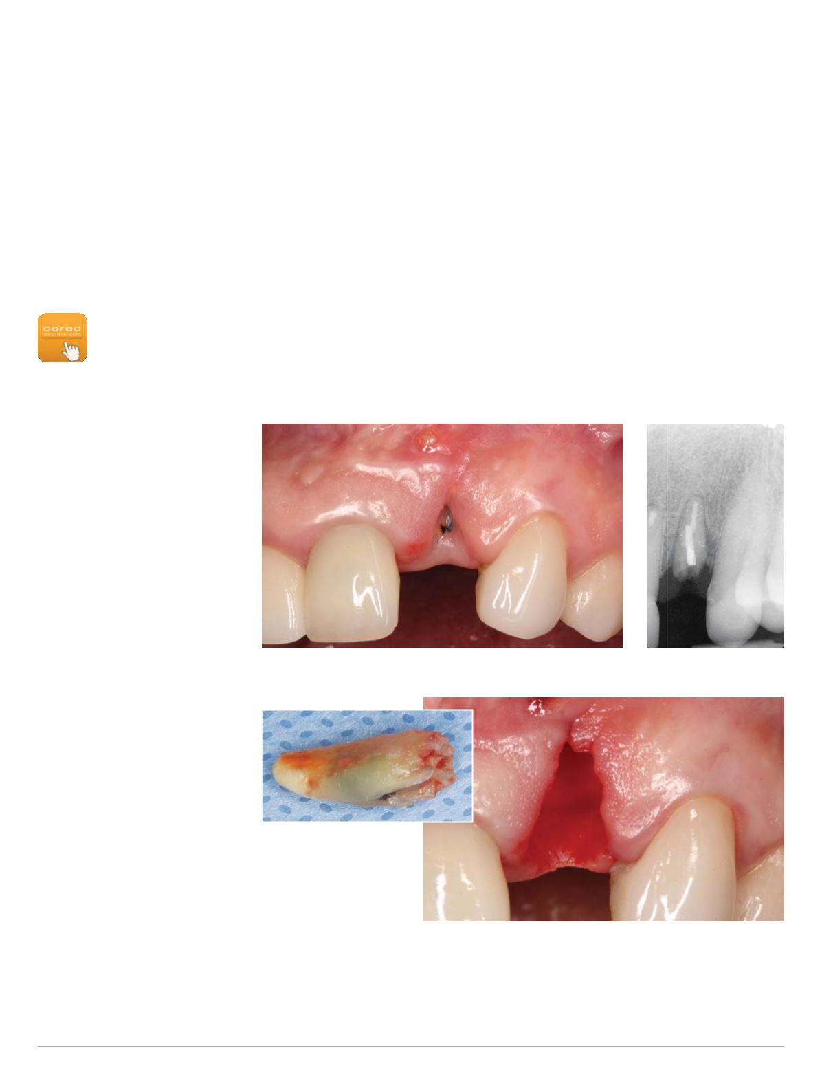
evaluation of a fractured tooth #10.
The initial clinical and periapical
radiographic evaluation revealed a
fractured root tip in site #10 with a
combined soft and hard tissue defect
andmoderate facial gingival recession
(Figs. 1-2). This finding led to a hope-
less prognosis for this tooth and after
discussion of various treatment alter-
natives with the patient, the decision
was made to extract the tooth and
replace it with an implant-supported
restoration. A two-phase treatment
planwas devised consisting of extrac-
tion of the root tip #10 with simul-
taneous hard and soft tissue recon-
struction to be followed by implant
placement in site#10 sixmonths later.
The initial surgical procedure was
performed under local anesthesia
and intravenous conscious seda-
tion. In addition, venous blood was
collected to obtain a Platelet Rich
Fibrin (PRF) concentrate via a centri-
fuge system. The remaining root tip in site #10 was extracted and the
extraction socket was thoroughly debrided revealing the extent of the
significant hard and soft tissue defect (Figs. 3-4). Subperiosteal buccal
and palatal pouches were prepared in site #10 without any vertical
Figs. 1-2: Preoperative clinical view (left) and periapical radiograph (right) of fractured tooth #10
Fig. 3: Extracted root tip
#10 (above)
Fig. 4: Post-extraction
clinical view of site #10
(right)
releasing incisions. A pedunculated connective tissue graft was then
harvested from sites #10-14 palatally taking care to leave the anterior
portion of the graft attached to ensure ongoing blood supply to the
graft (Fig. 5). This connective tissue graft was rotated and draped over


