49
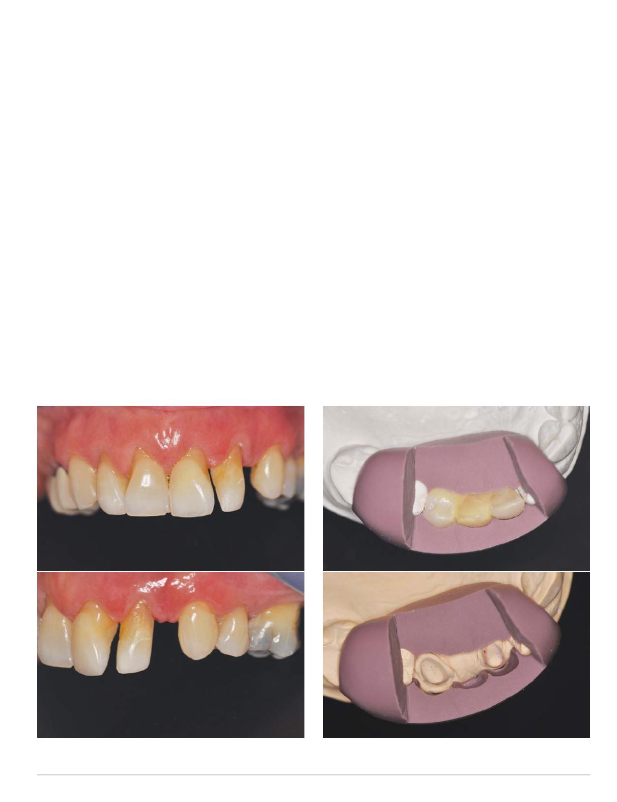
Fig. 3 Composite removed, #s 10, 11
Fig. 4 Jig relation of pre-op model and preparation
lateral function of the mandibular teeth led to somewhat regular
chipping and wear of the composite restorations.
It was also noted that the gum tissue surrounding tooth #10
looked significantly swollen and slightly darker in color. This
was most likely due to gingiva inflammation caused by the food
packing in the area caused by the poor contours of the composite
restoration.
Several treatment options were explored with the patient to
correct the tooth alignment. These included orthodontic treat-
ment to align the dentition followed by the placement of an
implant to create a tooth for the expected space remaining after
orthodontic treatment. This exceeded the time the patient was
willing to invest in a treatment solution, so more immediate
options were explored. A diagnostic model was fabricated and an
effort made to evaluate existing tooth dimensions in an attempt
to create a prototype of tooth dimensions that would blend better
with the contralateral teeth (Fig. 2).
It was not possible to create a prototype that replicated similar
tooth dimensions and proportions of the contralateral side strictly
by over-contouring existing teeth #10 and #11. The option that
provided the optimummatch of tooth contour was to add an addi-
tional tooth between #10 and #11. By removing the composite
restorations, it became clear that the mesiodistal width of both the
existing #10 and #11 allowed enough space for another tooth to be
added (Fig. 3). More control of the tooth dimensions was afforded
by using a three-unit fixed partial denture (FPD) prototype. The
need to limit the width of the tooth abutments led to broad prox-
imal contacts for the pontic and ensured sufficient size for the
connectors using a full contour e.max CAD FPD. A putty matrix
was fabricated from the diagnostic prototype to verify sufficient
space was created in the tooth preparations (Fig. 4). The proto-
type and plan was shared and discussed with the patient, and they
were accepted as the course of treatment.
A limited occlusal adjustment was done to create lateral guid-
ance on teeth #11-#12 in left lateral excursions to be reproduced
in the final FPD. This was done to provide some measure of stress
relief to #11 that would be abutment for the FPD. Teeth #10 and
#11 were prepared for all-ceramic abutment crowns taking care to


