52
|
CERECDOCTORS.COM
|
QUARTER 1
|
2016
to the observer, and accounted for more natural tooth proportions.
Also, the dark triangle caused by the slight tooth rotation and
gingival recession on the mesial of tooth #10 was minimized with
the CAD/CAM design. The hyperkeratinized area on the papilla
mesial to tooth #10 will continue to be monitored.
CAD/CAM fabrication of fixed partial dentures requires addi-
tional time compared to single crowns, with the potential for a
second appointment due to the extended times for milling and
processing the FPD. This may be a limiting factor for some clini-
cians to attempt full-contour CAD/CAM FPDs. However, the
level of chairside customization that can be achieved can easily
justify the additional time as it offers optimum control of the final
contours and shade matching.
For questions and more information, Dr. Fasbinder can be reached
at
| | |
VALCANAIA, NEIVA, FASBINDER
for 60 seconds. The tooth preparations were isolated with an
Isolite and selectively etched with 37 percent phosphoric acid for
20 seconds, rinsed and dried. Scotchbond Universal Adhesive was
scrubbed for 20 seconds on the preparations, dried until no move-
ment of the adhesive was visible, and RelyX Ultimate (3M ESPE)
A1 shade was used to adhesively lute the FPD with 60 seconds
VLC for each abutment. The occlusion was verified, and adjusted
areas re-polished intraorally with diamond-impregnated rubber
polishers (e.max polishers/Brassler) and diamond polishing paste.
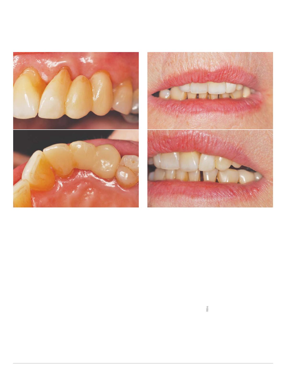
Post-operative evaluation of the e.max CAD FPD demonstrated
that the tooth proportions were significantly improved by the
addition of the pontic rather than over-contouring the two teeth.
A facial view of the case shows the natural transition between the
FPDand the patient’s dentition. Thiswas evenmore obviouswhen
observing lip positioning (Fig. 13). Even though the patient ended
up with one additional tooth on the left side, this was not obvious
Fig. 12 Restoration characterized try-in
Fig. 13 Final seat


