59
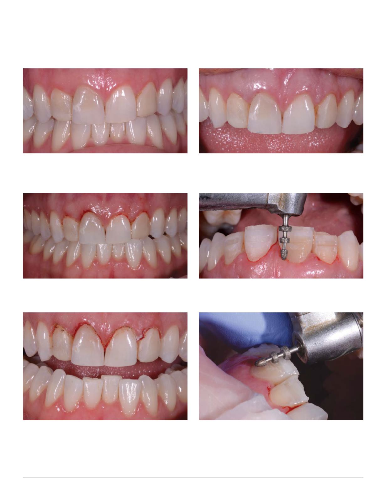
Fig. 1: Retracted preoperative view of a female patient unhappy with
the appearance of her smile, particularly the darkening tooth #8
and previously placed composite restorations
Fig. 2: With only minor gingival laser contouring to the cervical area
of tooth #8, the appearance and shape of the tooth were improved
Fig. 3: Immediate post-gingival contouring view of teeth #7 through
#10 demonstrating an enhanced height-to-width ratio
Fig. 4: The patient was allowed to heal for three weeks, resulting in
exceptional contours and enhanced tooth shape and length
Fig. 5: Once anesthesia had taken affect, initial depth cuts were made
using the RWMax .5/.7/.9 bur
Fig. 6: The three reduction planes for an anterior restoration were
properly completed


